Wide Mast Cell Tumor Resection with Sentinel (Axillary) Lymph Node Mapping and Excision
Signalment: 5.5-year-old, FS, Chinese shar-pei
History:
5-week history of a cutaneous mass on the right lateral thorax. The mass was pruritic.
Physical exam findings:
37.0 mm x 43.5 mm cutaneous, alopecic mass caudal to the right axilla
Axillary and superficial cervical lymph nodes normal size
Diagnostic and clinical staging tests:
CBC: no abnormalities
Serum no abnormalities
Cytology: high-grade MCT
Abdominal ultrasound: no significant abnormalities
Treatment:
Sentinel lymph node mapping and excision; and wide MCT resection and primary closure of the defect.
Outcome:
High grade III MCT (mitotic count 10/10 hpf) with complete histologic excision (lateral margins of 4.7 mm to 13.0 mm, and deep margins on 12.0 mm), and HN2 lymph node (i.e., early metastasis)
Complications:
None
Notes: The prognosis for this dog is unknown in some respects.
In the initial study reporting on the low/high grading scheme for MCTs, a median survival time of only 4 months was reported following surgical resection; however, this median survival time tripled when surgery was combined with chemotherapy.
More recent studies investigating the outcome following surgery alone for dogs with non-metastatic, high grade MCTs have shown more promising median survival times of 545 days, 899 days, and 1,046 days, with 1- and 2-year survival rates of 79% and 73%, respectively, in the latter study.
Two studies have shown no significant different in outcomes for dogs with HN2 (early) lymph node metastasis when treated with surgery alone versus surgery and chemotherapy; however, the majority of these dogs had high-grade II MCTs and not grade III MCTs.
So, what do we do with high-grade III MCTs with HN2 lymph node metastasis?
This dog is unfortunately a breed predisposed to high grade MCTs. Unfortunately, she developed a second mass on the right lateral flank (distant to the surgery site) within 1 month of surgery, and this was cytologically confirmed as a MCT. She is currently being treated with Palladia by our medical oncology team.
Tags: #MCT #SLN #SLNmapping

Preoperative sentinel lymph node (SLN) mapping radiograph following 4-quadrant, peritumoral injection of 0.5 ml of Omnipaque in each quadrant. While not as easy to detect as some SLN mapping procedures, there is contrast enhancement of the ipsilateral axillary lymph node (arrow).

Intraoperative SLN mapping with 4-quadrant, peritumoral injection of 0.5 ml sterile methylene blue in each quadrant. I typically do these injections on the border of the MCT but some surgeons prefer to do these injections 5-10 mm away from the MCT to minimize MCT degranulation and the effects that this may have on surgical margins.

Intraoperative SLN mapping with 4-quadrant, peritumoral injection of 0.5 ml sterile methylene blue in each quadrant. I typically do these injections on the border of the MCT but some surgeons prefer to do these injections 5-10 mm away from the MCT to minimize MCT degranulation and the effects that this may have on surgical margins.

Intraoperative SLN mapping with 4-quadrant, peritumoral injection of 0.5 ml sterile methylene blue in each quadrant. I typically do these injections on the border of the MCT but some surgeons prefer to do these injections 5-10 mm away from the MCT to minimize MCT degranulation and the effects that this may have on surgical margins.

Intraoperative SLN mapping with 4-quadrant, peritumoral injection of 0.5 ml sterile methylene blue in each quadrant. I typically do these injections on the border of the MCT but some surgeons prefer to do these injections 5-10 mm away from the MCT to minimize MCT degranulation and the effects that this may have on surgical margins.

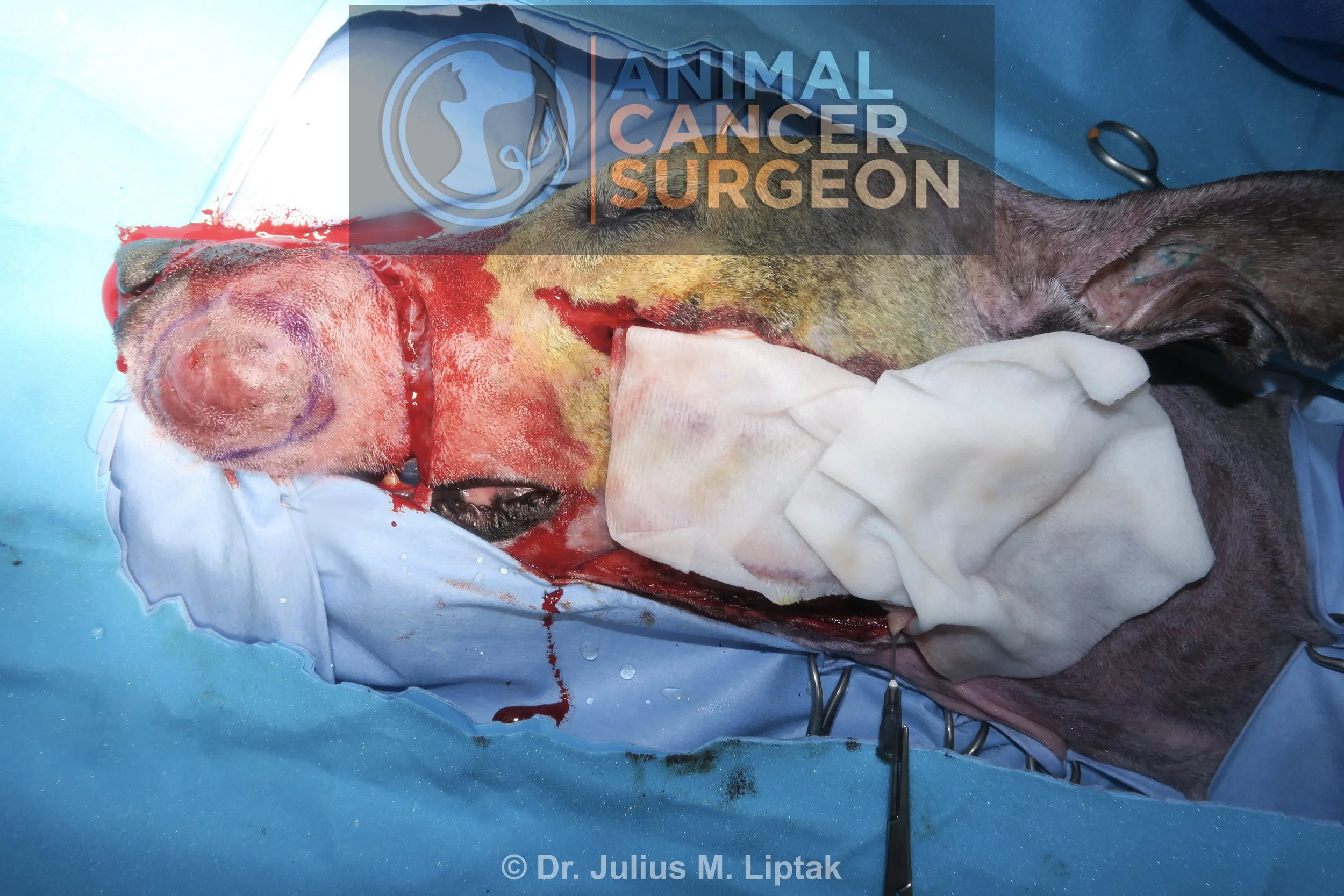
Following resection of the MCT with 2 cm lateral margins and part of the latissimus dorsi muscle for deep margins, blue coloured lymphatics could be seen heading in the direction of the axilla (see middle finger of assistant).

Once the blue coloured sentinel (axillary) lymph node was identified, this was excised with a LigaSure.

Immediate postoperative image following primary closure of the surgery site.