Lip Resection and Facial Axial Pattern Flap Reconstruction with Sentinel Lymph Node Mapping and Excision in a Dog with a High-Grade Mast Cell Tumor
Signalment: 10-year-old, FS, greyhound
History:
This dog originally presented to our medical oncology service in mid February 2025 with a 2-month history of a left rostral lip mass. A mast cell tumor (MCT) was diagnosed on fine-needle aspirate cytology, and a low grade MCT (mitotic count 0/10 hpf) was diagnosed histologically following a punch biopsy. She was transferred to our surgery service 5 weeks later for discussion on further staging (with sentinel lymph node mapping) and surgical options.
Physical exam findings:
30.5 mm x 38.9 mm left rostral lip mass
Mandibular lymph nodes normal size bilaterally
Diagnostic and clinical staging tests:
CBC: mild thrombocytopenia
Serum biochemistry: mildly increased creatinine with high normal BUN and SDMA
Cytology: MCT
Histopathology: low grade MCT with a mitotic count of 0/10 hpf
Treatment:
Sentinel lymph node mapping and excision; and wide MCT resection with lip reconstruction using a facial axial pattern flap.
Note: I personally prefer the term “facial axial pattern flap” for this flap but it is also called an angular oris axial pattern flap. I prefer facial axial pattern flap as the direct arterial supply is not solely the angular oris artery but also the superior and inferior labial arteries, all of which are separate branches of the facial artery.
Outcome:
High grade III MCT (mitotic count 17/10 hpf) with complete histologic excision (lateral margins of 0.7 mm to 16.0 mm, and deep margins on 9.8 mm), and HN0 lymph node (i.e., no metastasis)
Complications:
Flap edema. This is a common complication with the facial axial pattern flap, occurring in 89% of dogs and 100% of cats.
Distal flap necrosis and partial incisional dehiscence. While this is less common than edema, partial incisional dehiscence and distal flap necrosis are reported in 30% and 22% of dogs, respectively. I was concerned about the possibility of distal flap necrosis because of the conformation of the head of this dog; she had a long nose and a relatively short lateral face, which, combined with a rostral lip defect, meant that the flap dimensions were relatively smaller and this needed to be transposed a greater distance becaiuse of the length of the nose and the rostral location of the defect. The distal flap necrosis was debrided and managed as an open wound, resulting in full thickness loss of the rostral 50% of the flap/lip.
Surgical site infection
Notes: In one study investigating the accuracy of pre-treatment biopsy in dogs with MCTs (Shaw et al, Vet Comp Oncol, 2018), the authors found an overall concordance rate of 96% based on the Patnaik grading system, and an overall concordance rate of 92% based on the Kiupel grading system. The accuracy of the various biopsy techniques (wedge, punch and needle core) when compared with excisional biopsy was 92%, 100% and 100%, respectively, based on the Patnaik grading system, and 90%, 95% and 100%, respectively, based on the Kiupel grading system. Of the cases with discordant results, the pre-treatment biopsies tended to underestimate the grade of the tumor. The authors concluded that pre-treatment biopsies are sufficiently accurate for differentiating low-grade from high-grade MCTs, regardless of biopsy technique or tumor location.
Obviously, while this is generally true, the preoperative biopsy was not accurate in this particular dog considering a low grade MCT with a mitotic count of 0/10 hpf was diagnosed preoperatively and it was definitively diagnosed with a high-grade III MCT with a mitotic count of 17/10 hpf postoperatively. The latter is more consistent with MCTs in a perioral or muzzle location as the majority of these have a higher histologic grades with a more aggressive biological behaviour. In this case, there was no evidence of lymph node metastasis, so the prognosis should be good for this dog. Studies investigating the outcome following surgery alone for dogs with non-metastatic, high grade MCTs have shown more promising median survival times of 545 days, 899 days, and 1,046 days, with 1- and 2-year survival rates of 79% and 73%, respectively, in the latter study.
Tags: #MCT #SLN #SLNmapping #facialaxialpatternflap #reconstruction #distalflapnecrosis

Sentinel lymph node mapping was performed with four-quadrant, peritumoral injection of 2 ml Omnipaque. Radiographs taken at 1 min show contrast enhancement of the ipsilateral medial and lateral mandibular lymph nodes (arrows). Note the contrast-enhanced lymphatics draining into these lymph nodes.

Sentinel lymph node mapping was performed with four-quadrant, peritumoral injection of 2 ml Omnipaque. Radiographs taken at 1 min show contrast enhancement of the ipsilateral mandibular lymph nodes (arrow).

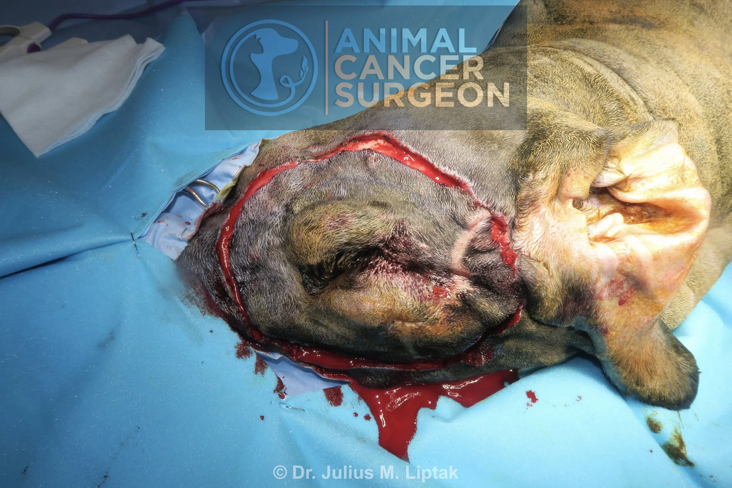
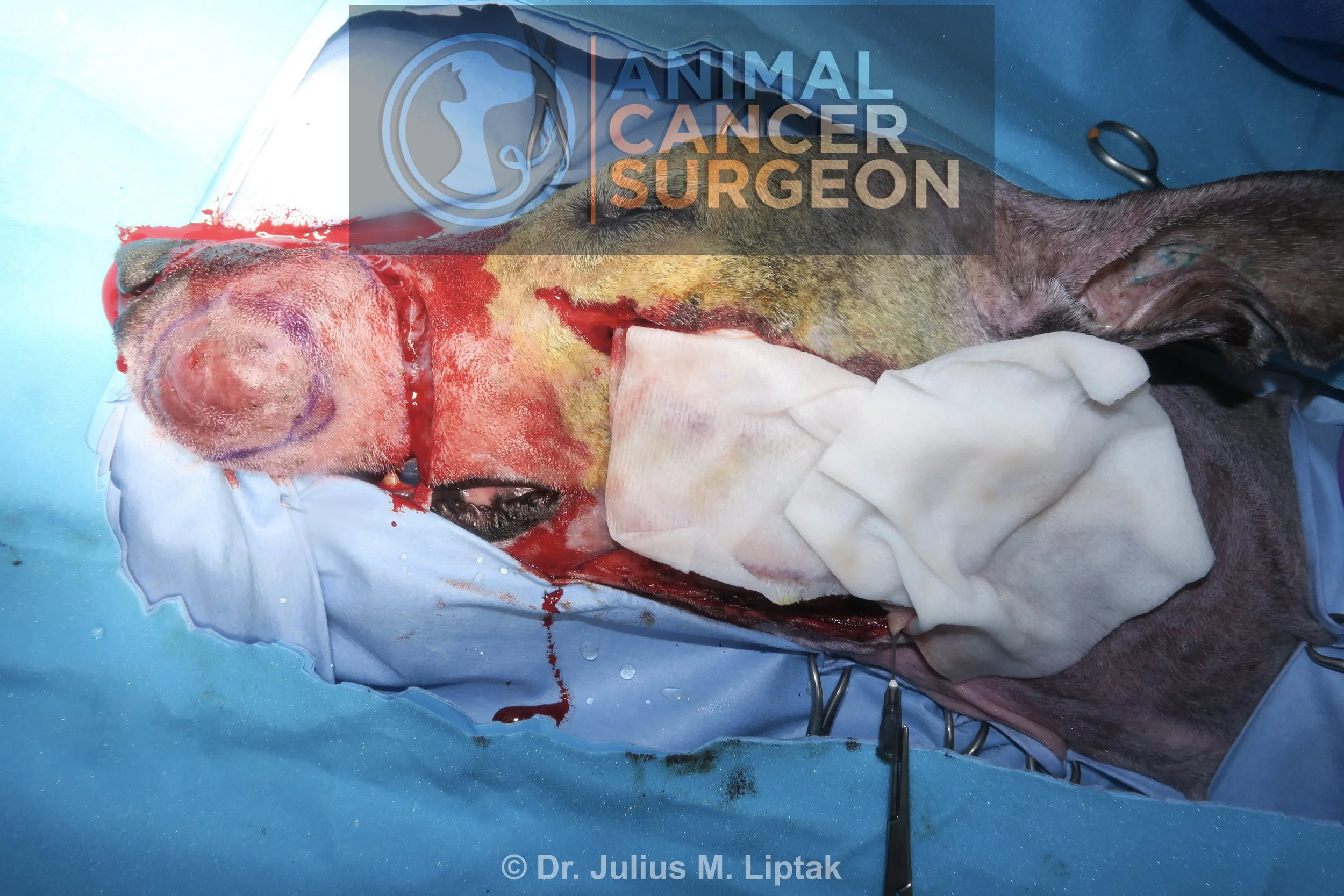
The margins of the tumor, 2 cm lateral margins around the MCT, and the borders of the facial axial pattern flap were marked with a sterile marker pen.

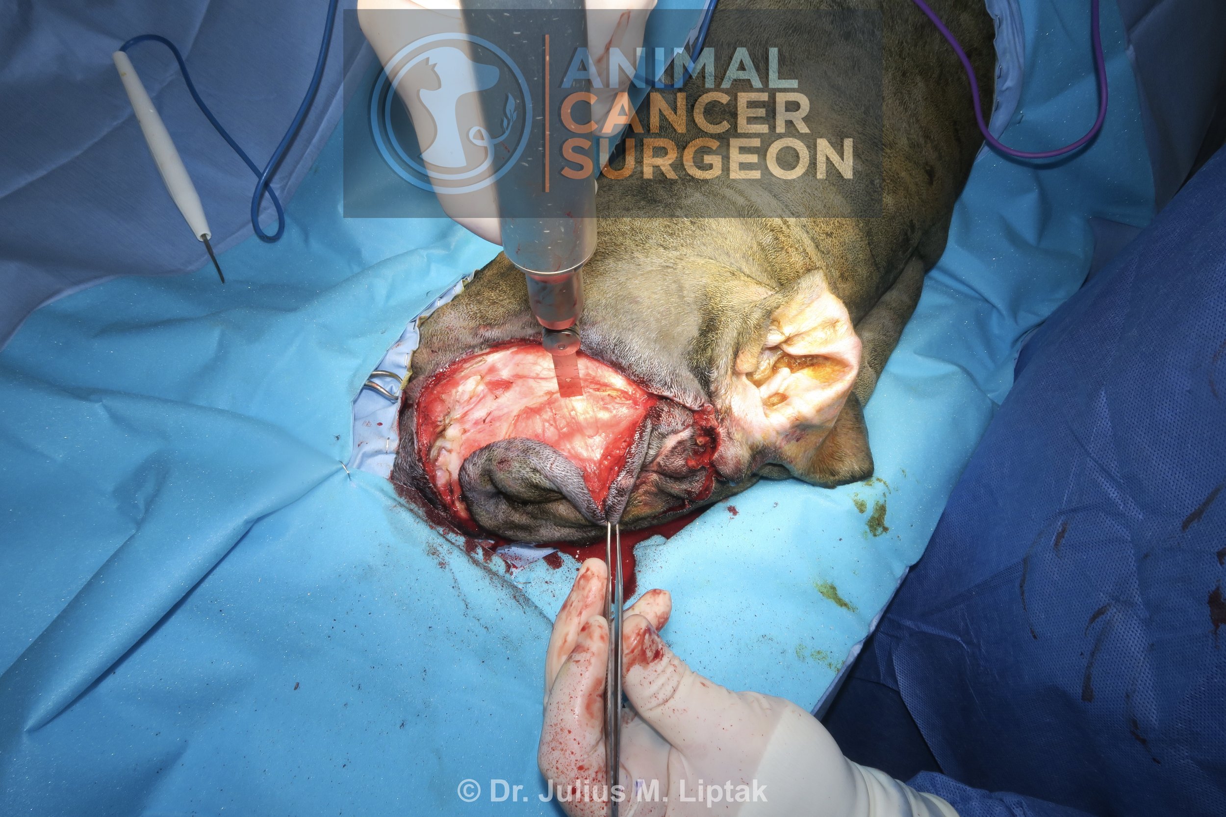
The facial axial pattern flap was raised before resection of the lip MCT. The borders for this flap are along the ventral aspect of the zygomatic arch dorsally, along the ventral aspect of the mandible ventrally, and then these two incisions are connected caudally at the level of external ear canal. You can appreciate how short this flap is relative to the length of the nose.

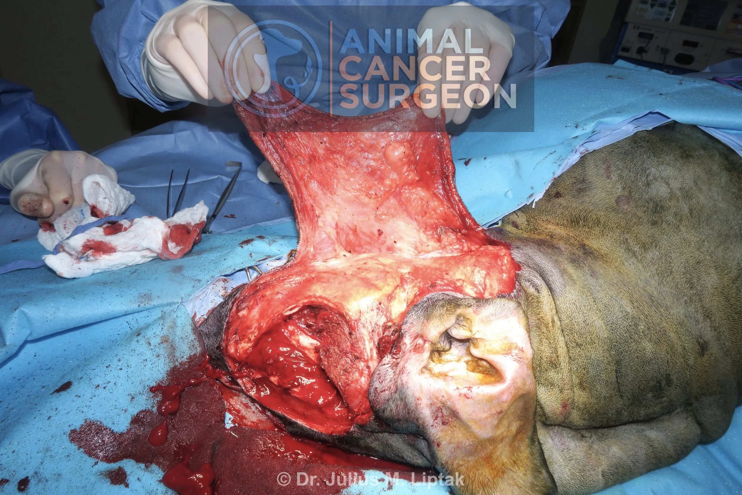
The flap was undermined below the level of the panniculus muscle, preserving the subdermal plexus. The mandibular lymph nodes were visible after the flap was raised.

The medial and lateral mandibular lymph nodes were extirpated using a LigaSure. Note that intraoperative sentinel lymph node mapping was not used because we knew that the mandibular lymph nodes would be well exposed with this approach.

The facial axial pattern flap after it was completely raised.

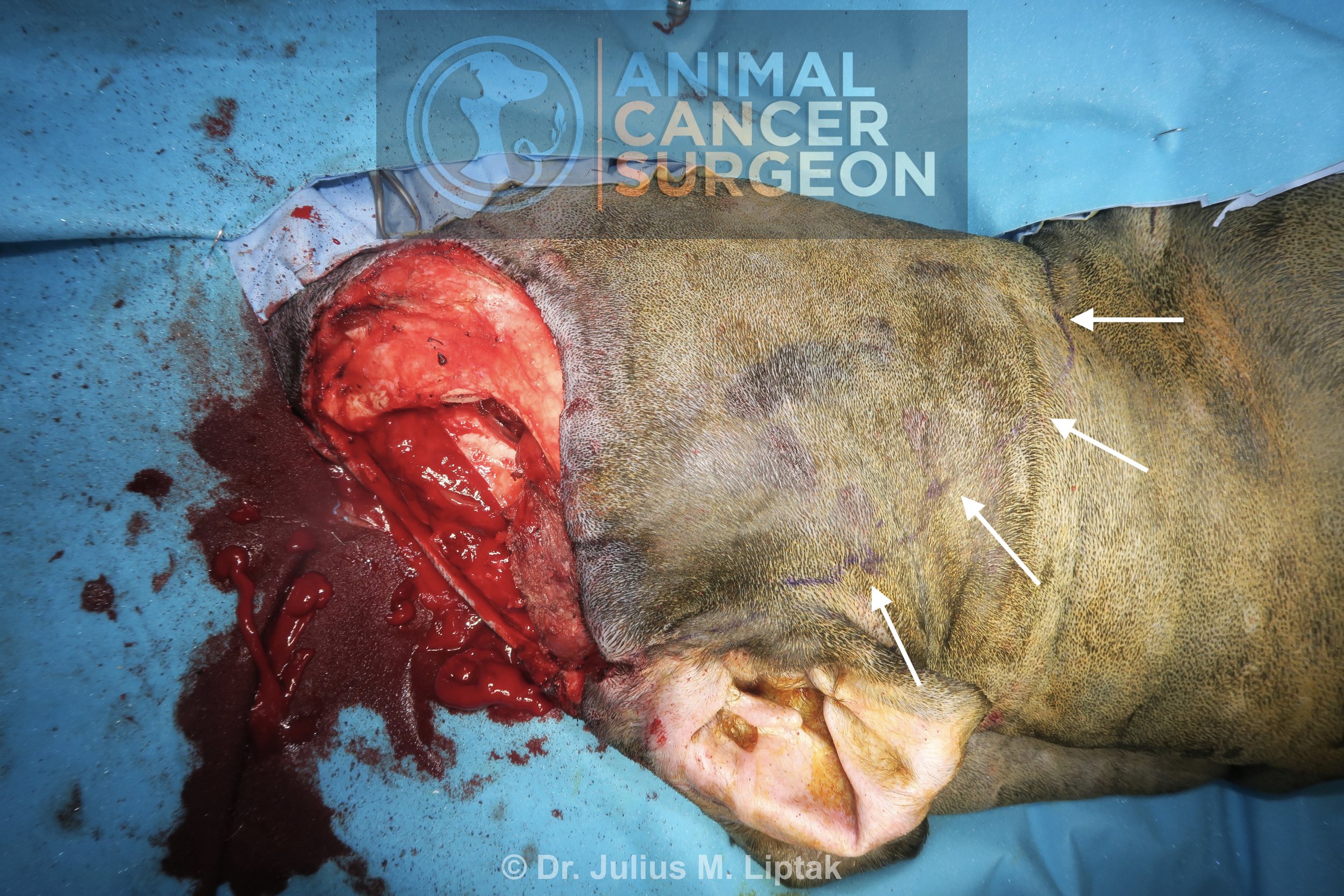
I was feeling a little more confident after transposing the facial axial pattern flap into the likely defect, as can be seen with the rostral extent of the flap in this image.
The flap was protected with moistened gauze swabs, and the lip MCT was resected with 2 cm lateral margins and full thickness lip for deep margins.

Gross appearance after full thickness resection of the lip MCT with 2 cm lateral margins.

The rostroventral corner of the flap was sutured initially. Note that haired lip will now form the medial aspect of the reconstructed lip.

The facial axial pattern flap was then sutured to the residual labial mucosal margin to complete the intraoral closure.

The lateral aspect of the flap was then sutured into position. The tension was highest on the rostral aspect of the flap, as well as the rostral aspect of the donor site closure; both of these areas eventually underwent necrosis and dehiscence.

Appearance of the facial axial pattern flap 1 day after surgery. Notice the edema and swelling in the flap, which is common in facial axial pattern flaps (occurring in 89% of dogs).

8 days after surgery, the area of the flap indicated by the arrows is slightly darker and has a more leathery feel, indicating likely distal flap necrosis.

The area of distal flap necrosis extended after a further 4 days (12 days postop) (arrows). This was debrided, cultured, and treated with empirical (and, later, culture-directed) antibiotics.

Gross appearance of the dog 14 days after her original surgery, and 2 days after her debridement. Her open wound continues to heal by second intention and, while cosmesis is less than desirable, she is functioning completely normal and has returned to her normal quality of life.