Filtering by Tag: orbitectomy

Orbitectomy for a Dog with a Grade I Multilobular Osteochondrosarcoma

Signalment: 3.5-year-old, FS, golden retriever

History:

This dog was referred after a 2-month history of right-sided conjunctivitis and increased intraocular pressure (21-25 mm Hg) progressing to exophthalmos with an inability to retropulse the right eye. A CT scan was performed which showed a well-circumscribed osseous mass arising from the medial orbital bone.

Physical exam findings:

  • Mild to moderate right-sided exophthalmos

  • Inability to retropulse right eye

Diagnostic and clinical staging tests:

  • CBC: no abnormalities

  • Serum biochemistry: no abnormalities

  • CT scan: 3.2 cm x 3.4 cm x 3.9 cm osseous mass arising from the medial aspect of the right orbit, with mild enlargement of the right medial retropharyngeal lymph node. No evidence of lung metastasis.

Treatment:

Orbitectomy combined with resection of the zygomatic arch and coronoid process of the mandible.

Outcome:

  • Grade I multilobular osteochondrosarcoma (MLO) with complete excision (bone margins: 20.0 mm to 30.0 mm; deep margins: 11.0 mm)

  • Complications:

    • Blindness (OD), immediate. This was expected to some degree because of prolonged retraction on the optic nerve and vessels to expose the bone margins for resection of the mass.

    • Sneezing and difficulty opening jaw, 6 weeks postop. The cause for these remain undiagnosed and unresolved. While local tumor recurrence is a possibility, I think this is highly unlikely considering that this tumor was completely excised with very good surgical and histologic margins, and that this is a grade I MLO (which are typically slow growing tumors). A repeat CT has been recommended but this has not been done yet.

Video link: https://www.youtube.com/watch?v=eBrdiKihhZA&t=370s

Tags: #MLO #orbitectomy

Preoperative CT scan showing a large osseous mass arising from the medial aspect of the right orbit.

Preoperative CT scan showing a large osseous mass arising from the medial aspect of the right orbit. Note that this mass extends to the orbital bone overlying the frontal lobe of the brain.

3D reconstruction showing the large orbital mass. These reconstructions are very helpful in surgical planning, especially the approach (in this case, zygomatic arch resection and resection of the coronoid process of the mandible) and the margins (in this case, knowing that we would see brain and hence use a more delicate instrument to make the osteotomies in this region).

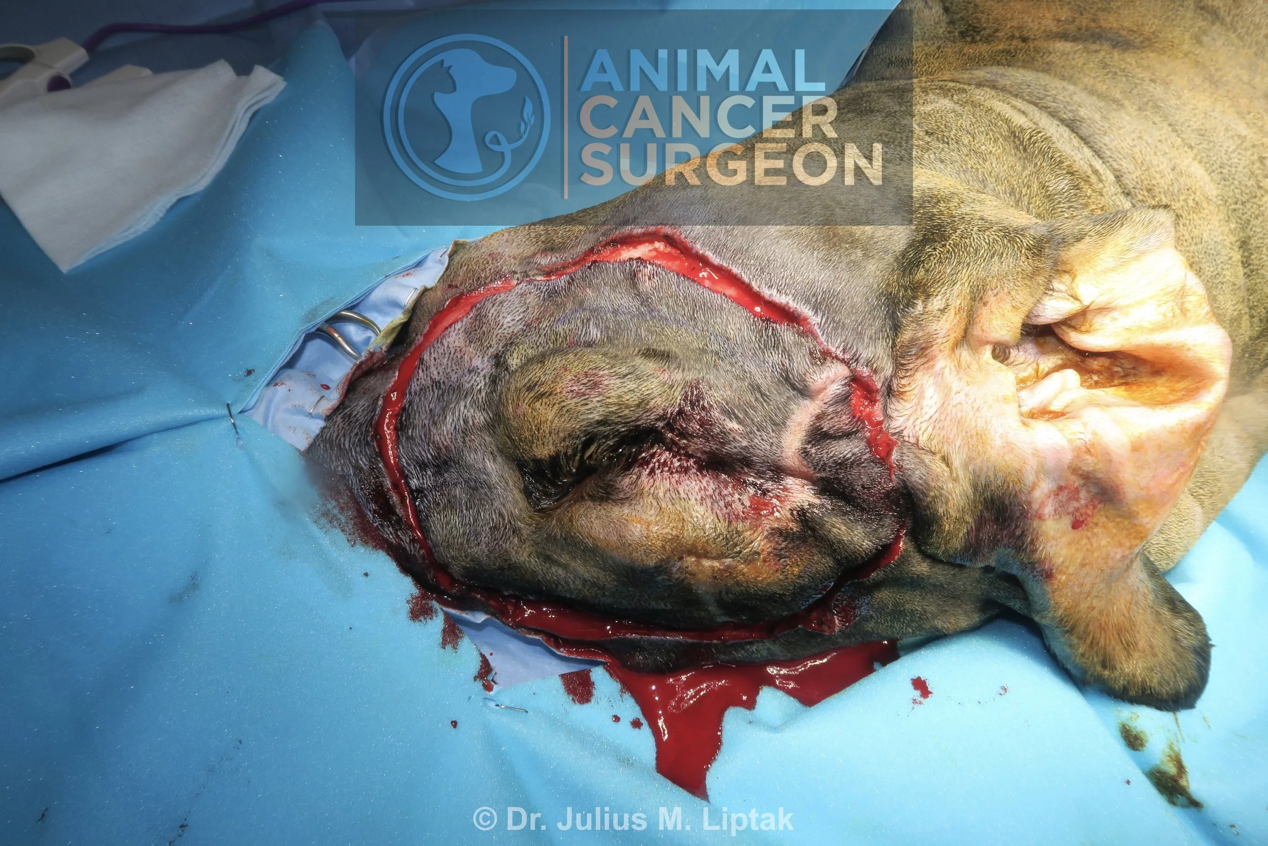
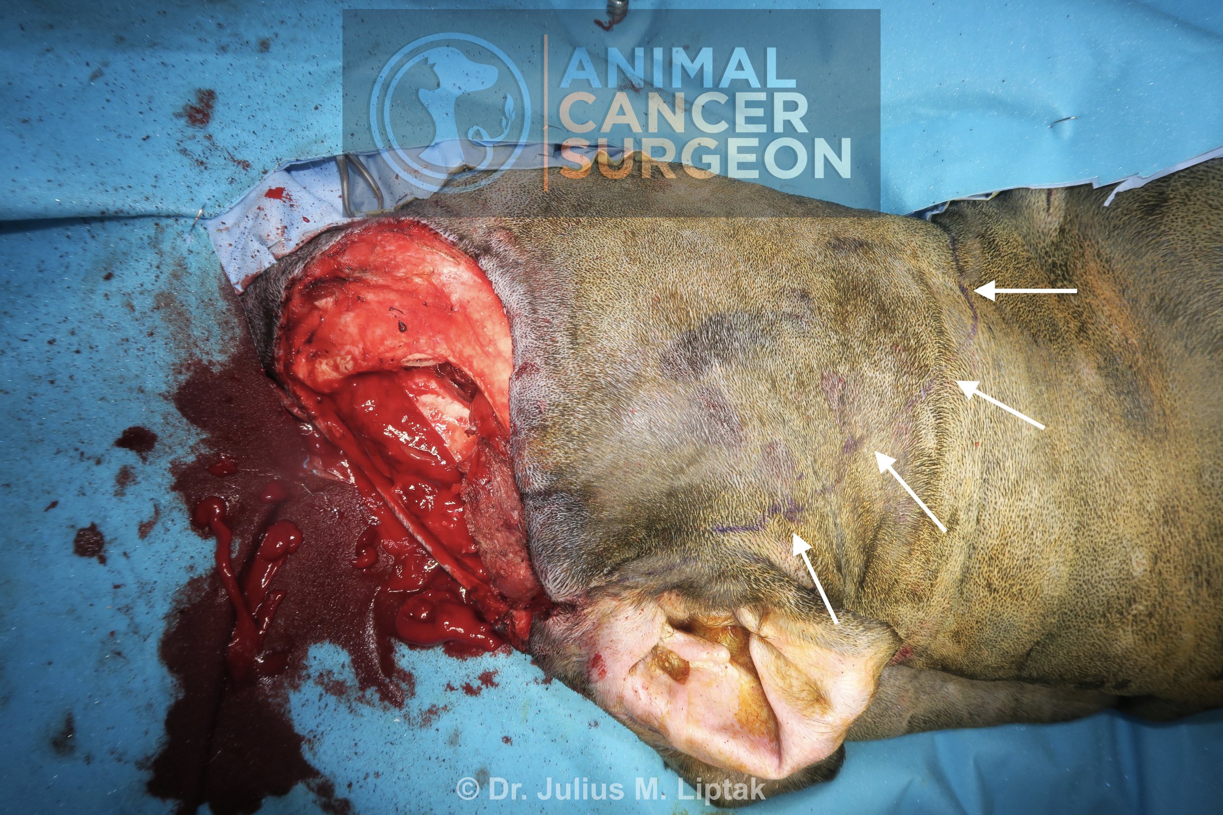
The initial surgical approach involved a skin incision along the zygomatic arch and extending on to the dorsolateral aspect of the caudal muzzle.

Electrosurgery was used to incise and elevate the temporal (pictured) and masseter muscles from the dorsal and ventral aspects of the zygomatic arch, respectively.

Soft tissues attached to the medial aspect of the zygomatic arch were freed using a periosteal elevator.

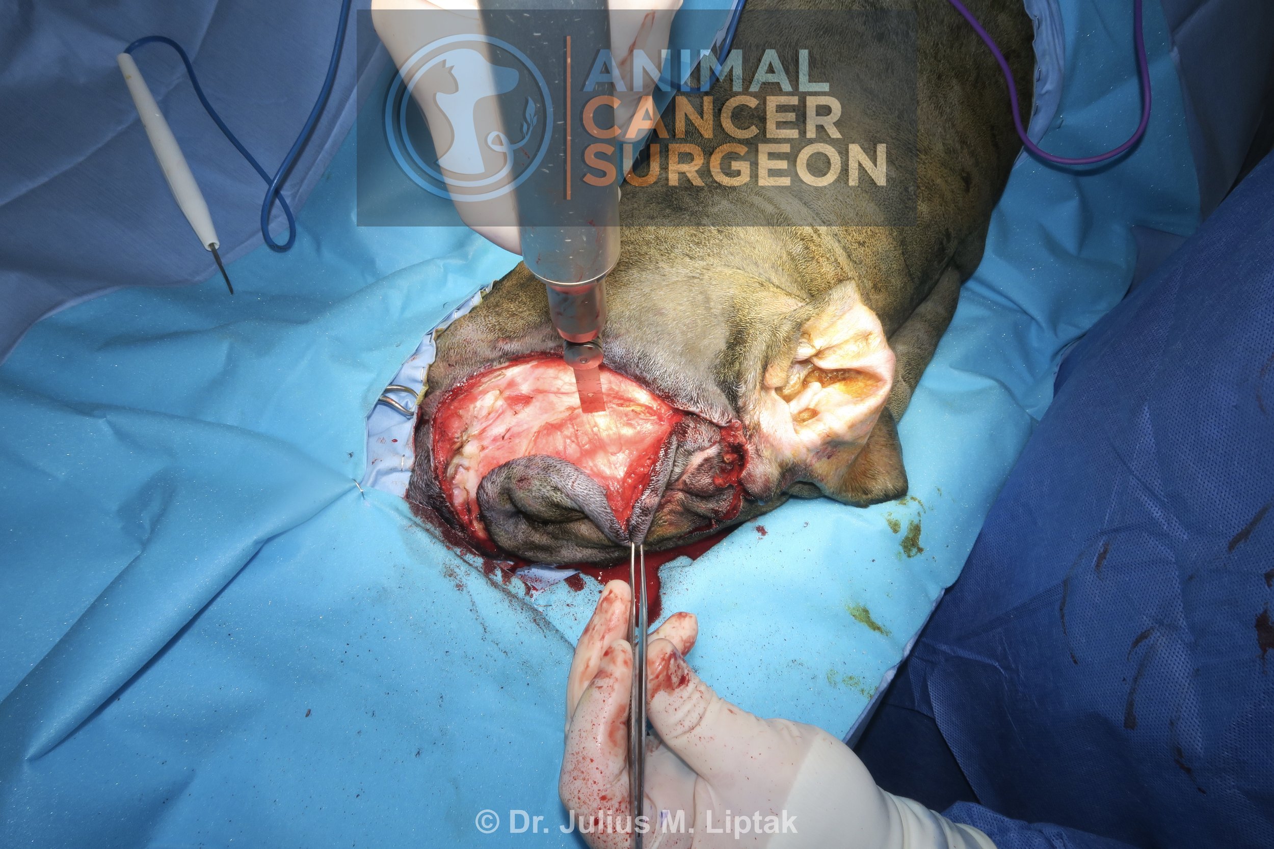
The first step to exposing the orbit is a zygomatic arch resection. Once the zygomatic arch is exposed and freed of all its soft tissue attachments, the caudal zygomatic bone osteotomy was performed with a sagittal saw. The periosteal elevator is positioned to protect the deeper soft tissues from iatrogenic trauma from the saw blade.

The rostral zygomatic arch resection was completed with a sagittal saw and osteotome and mallet. This osteotomy was done in such a way that a portion of the caudal maxilla was included in the osteotomy, but dorsal to the teeth.

The completed zygomatic arch resection before removal of the zygomatic arch. I do not replace the zygomatic arch after surgery. There is no functional need for it and I very rarely see a cosmetic defect as a result of its absence.

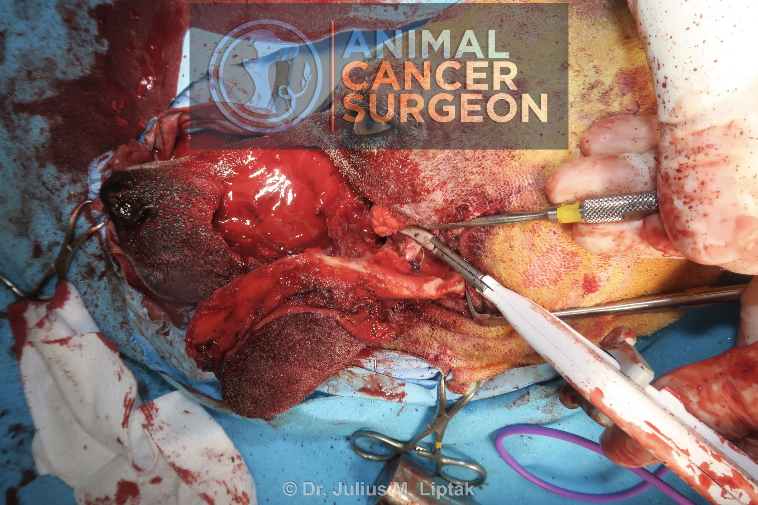
The orbit mass was easily exposed after dissecting through the superficial soft tissues; however, the caudal aspect of the mass and caudal orbit were difficult to visualize because of the vertical ramus of the mandible.

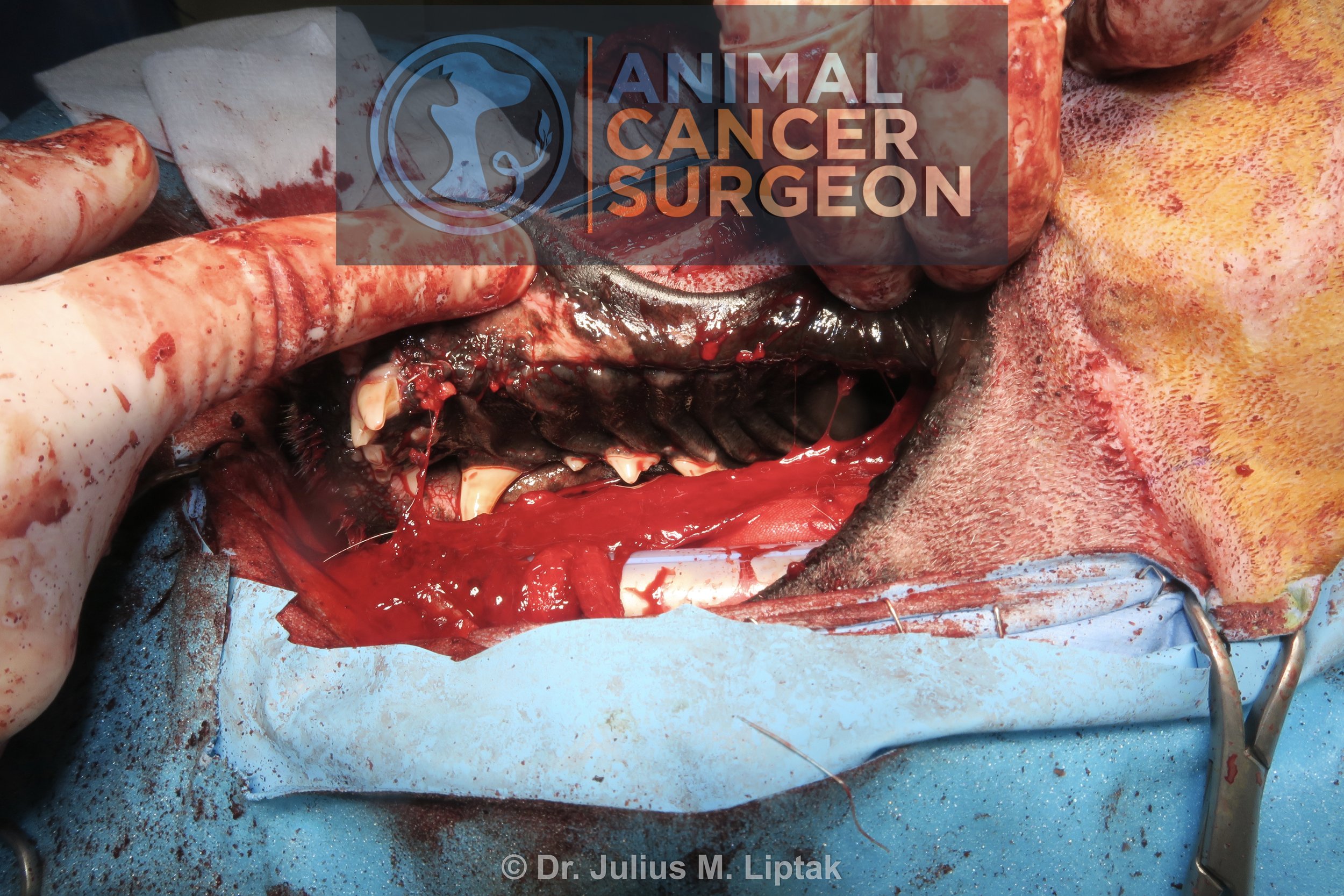
The coronoid process of the mandible was osteotomized with a sagittal saw dorsal to the temporomandibular joint to improve visualization of the caudal orbit.

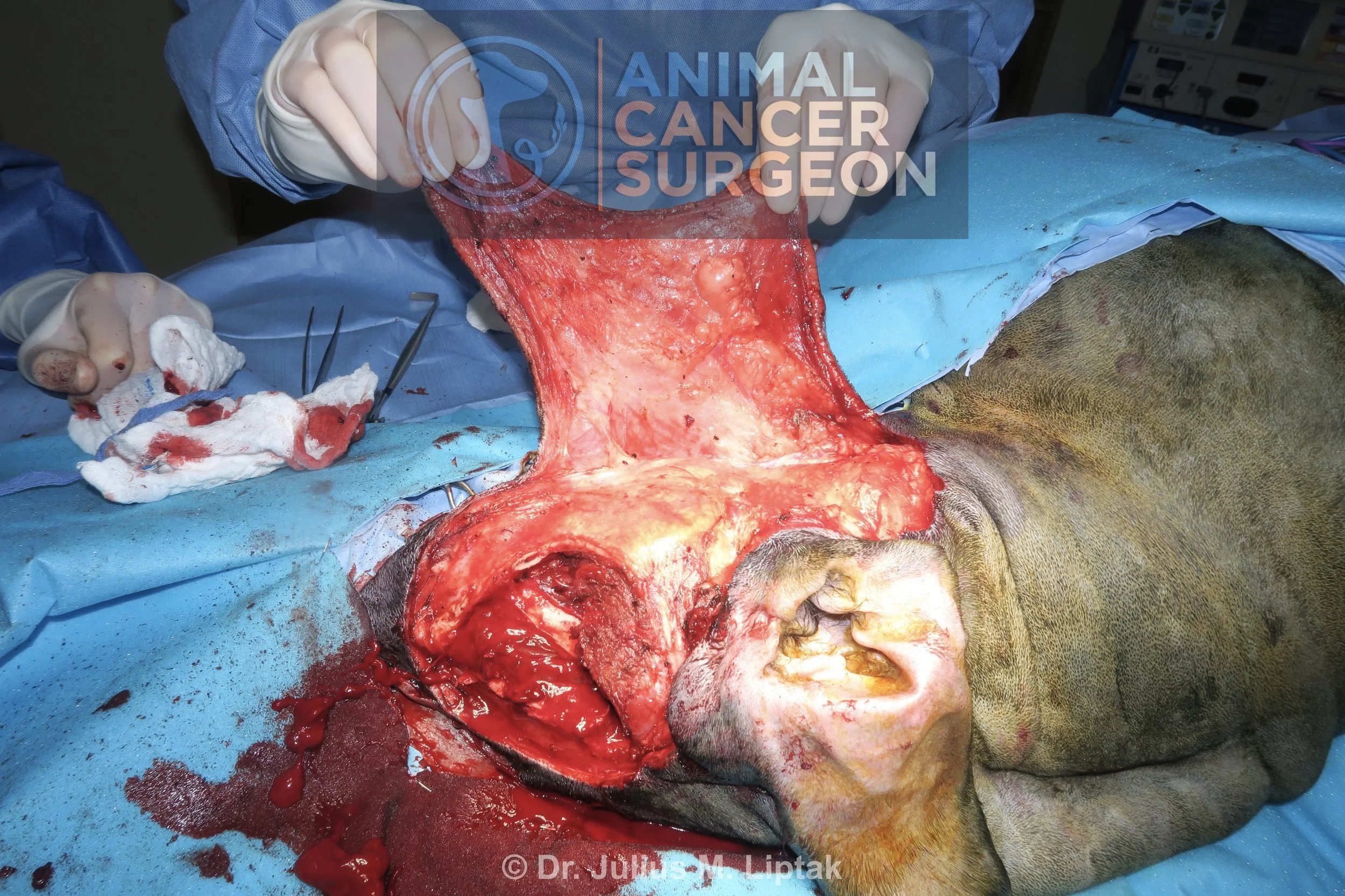
Following the removal of the coronoid process of the mandible, the exposure of the caudal orbit was markedly improved.

I wanted to save the eye, which we did anatomically, but the dog was blind in this eye after surgery. I suspect this was a result of protracted and marked retraction of the caudal globe structures (vessels and optic nerve) during resection of the mass. The majority of the dorsal osteotomy was done with a piezotome in an effort to reduce iatrogenic trauma to the underlying brain; however, while accurate and precise, a peizotome is a painfully slow technique to do osteotomies, even in thin bone such is in this region.

Intraoperative image of the orbitectomy site following removal of the orbital mass from the surgical field.

Bleeding from the meninges was controlled with hemostatic foam.

The wound was then closed primarily in three layers: temporal and masseter muscle fascia, subcutaneous, and skin.

Postoperative specimen image of the resected orbital mass. The mass had a small attachment to the orbital bone, which meant that bone margins were easier to achieve than if this had a broad-based attachment to the bone. Additional caudal margins were obtained because I was not satisfied with my original margins. Histologically, this was a complete excision with the narrowest histologically tumor-free margins being 11 mm.

Immediate postoperative CT scan showing the extent of the orbitectomy along the rostral aspect of the surgical field.

Immediate postoperative CT scan showing the extent of the orbitectomy along the caudal aspect of the surgical field at the level of the frontal lobe of the brain.

Reconstructed CT view of the orbitectomy.

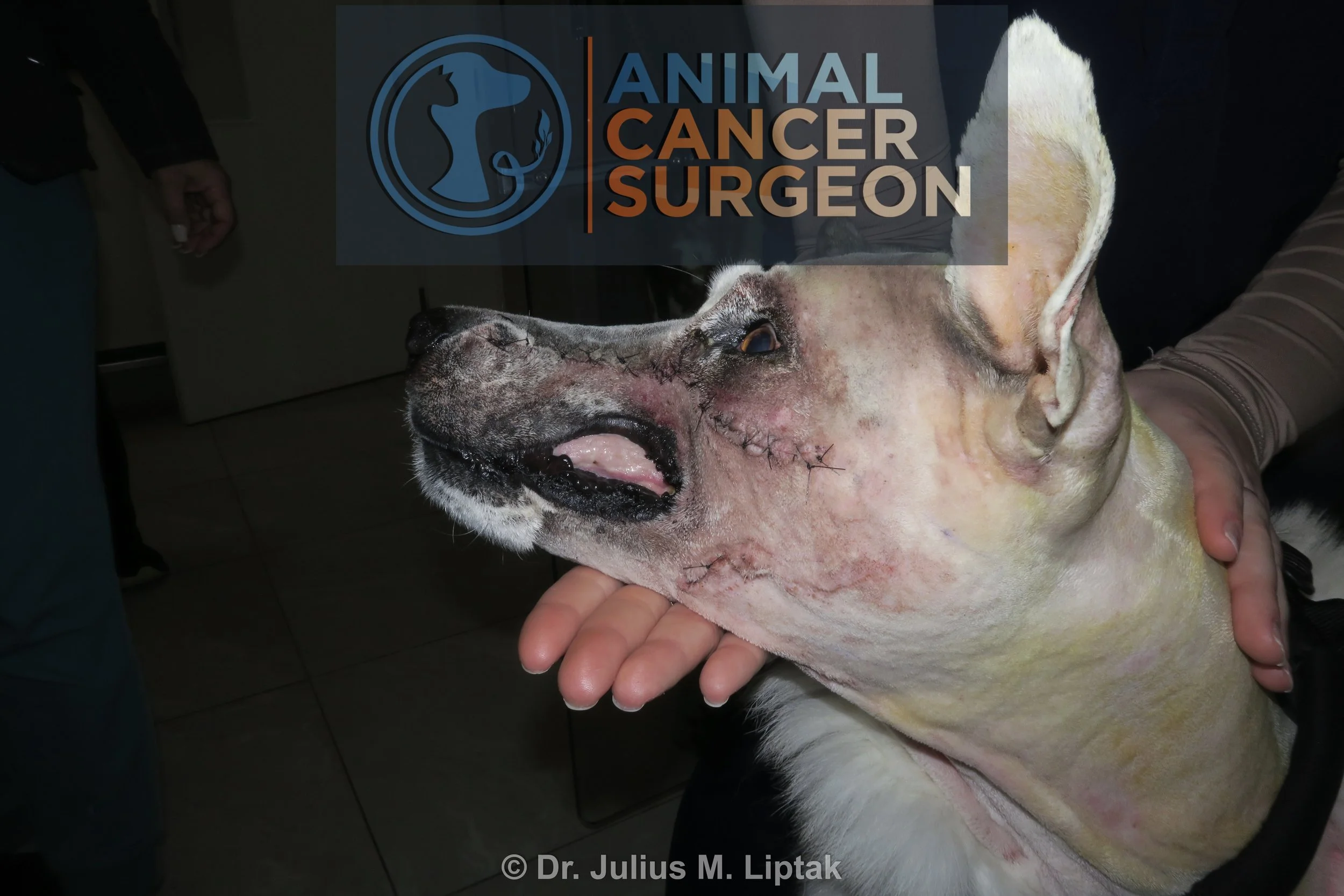
Day 1 postoperative appearance. This swelling and bruising took over 1 week to subside. The swelling made initial assessment of eyesight difficult, but the dog did not have a menace reflex initially and had not regained a menace reflex by week 6 after surgery.

Total Unilateral Maxillectomy via a Combined Approach for Resection of a Biologically High-Grade but Histologically Low-Grade Fibrosarcoma

Signalment: 7.5-year-old, MN Siberian husky

History:

This dog presented with a 1-month history of a left-sided muzzle mass. He did not have any other clinical signs associated with this mass.

Physical exam findings:

  • 43.5 mm x 50.4 mm firm and fixed mass extending along the left muzzle from rostral to the orbit to the rostral aspect of the muzzle

  • No involvement of the oral cavity

  • Mandibular lymph node normal size on palpation

Diagnostic and clinical staging tests:

  • CBC: no abnormalities

  • Serum biochemistry: mildly increased ALT, ALP, and GGT

  • Punch biopsy: sarcoma

  • CT scan: left-sided soft tissue mass extending along the lateral aspect of the muzzle. No evidence of either lymph node or lung metastasis.

Treatment:

Total unilateral maxillectomy via a combined dorsolateral skin and intraoral approach, including en bloc resection of the skin overlying the mass. A biologically high-grade but histologically low-grade fibrosarcoma was suspected based on its presentation and CT appearance, so 3 cm soft tissue and bone margins were used for resection, including a rostral orbitectomy. We had initially planned to reconstruct the soft tissue defect with a facial axial pattern flap; however, after resection, this was not required as we were able to preserve the ventral lip margin.

Outcome:

  • Hi-lo fibrosarcoma with complete excision (skin margins: 7.2 mm to 12.0 mm; bone margins: 6.4 mm to 20.0 mm)

  • No complications

Video link: https://www.youtube.com/watch?v=6bFNQldai0Q&t=1042s

Tags: #hiloFSA #FSA #orbitectomy #maxillectomy

Preoperative CT scan showing a soft tissue mass extending along the lateral aspect of the left muzzle. This is a more consistent presentation for a hi-lo fibrosarcoma than any other tumor type, and correlated with the preoperative biopsy results.

Preoperative CT scan showing a soft tissue mass extending along the lateral aspect of the left muzzle. There was a small amount of bone involvement (arrow), but otherwise this was entirely a soft tissue mass. This is a more consistent presentation for a hi-lo fibrosarcoma than any other tumor type, and correlated with the preoperative biopsy results.

While the skin overlying the mass was freely movable, this was resected en bloc with the mass because of the high likelihood that this was a hi-lo fibrosarcoma, which is associated with a high risk of local tumor recurrence. Note that the borders for a planned facial axial pattern flap have been marked with a sterile marker pen; however, we did not end up using this flap for reconstruction of the lip defect.

The soft tissue dissection is then continued deeply to the level of the maxilla and full thickness through the lips with 3 cm soft tissue margins in the subcutaneous space.

An intraoral incision is made along the gingival margin, connecting the skin incision to create a bipedicle flap (near my left thumb). Pictured is an incision being made along the medial aspect of the dental arcade into the mucoperiosteum of the hard palate.

The mucoperiosteum is then elevated and reflected medially to expose the palatine bone for its osteotomy.

A sagittal saw was used to osteotomize the rostral aspect of the zygomatic arch and caudal maxilla.

A sagittal saw was used to continue the osteotomy along the dorsal midline of the maxilla.

Once the dorsal osteotomy was completed, this was then continued rostrally between the left canine and corner incisor teeth.

The sagittal saw was then used to perform an osteotomy along the hard palate bone.

Finally, an osteotome and mallet were used for the osteotomy within the rostral aspect of the orbit, connecting the dorsal maxilla and palatine osteotomies. This is typically done with an osteotome and done last as this cut has the greatest potential for brisk bleeding from the maxillary artery. While techniques have been described to pre-ligate the maxillary artery, I rarely find this necessary.

Medial soft tissue attachments to the resected maxillary bone are then transected; in this case, with a LigaSure.

The resected maxillectomy segment immediately prior to being removed from the surgical field.

Appearance of the maxillary defect following removal of the maxillectomy segment for the surgical field.

The mucosa and submucosa of the lip was split to create a labial mucosal-submucosal flap, and then this was sutured to the submucosa of the mucoperiosteum. This is easier to do from the lateral skin incision.

The mucosa and submucosa of the lip was split to create a labial mucosal-submucosal flap, and then this was sutured to the submucosa of the mucoperiosteum. This is easier to do from the lateral skin incision.

Intraoral appearance of the palatine closure.

I had originally planned to close the lateral muzzle defect with a facial axial pattern flap; however, we were able to preserve the ventral aspect of the upper lip and close this defect primarily.

Primary closure of this defect resulted in elevation of the caudal aspect of the upper lip but I preferred this to the potential complications associated with flap reconstruction of this defect.

Postoperative specimen image of the maxillectomy, lateral aspect. This was confirmed as a hi-lo fibrosarcoma with the narrowest histologic tumor-free margins being 6.4 mm.

Postoperative specimen image of the maxillectomy, medial aspect. This was confirmed as a hi-lo fibrosarcoma with the narrowest histologic tumor-free margins being 6.4 mm.

Appearance of the dog 1 day postoperatively.

Subcutaneous Biologically High Grade and Histologically Low Grade Fibrosarcoma Resection with Orbitectomy and Rotation Flap Reconstruction

Signalment: 7-year-old, MN bernadoodle

History:

This dog initially presented to me in late November 2023 with a rapidly growing subcutaneous mass, measuring 36.2 mm x 58.4 mm, on the dorsal to right dorsolateral aspect of the head. An incisional biopsy of the mass was read out as a fibroma. Wide resection of the mass was done a week later; histopathology of the mass was consistent with an incompletely excised (on the deep margin) biologically high grade and histologically low grade (so called hi-lo) fibrosarcoma.

The dog presented for a recheck exam in mid-January 2025 because of concern for a local recurrence. There was a palpable mass in the region of the surgical scar and a subsequent incisional biopsy confirmed a hi-lo fibrosarcoma. Three days prior to his planned surgery, a second mass appeared along the ventral aspect of the right lower eyelid. This was not physically connected to the recurrent mass. While a biopsy of this mass was discussed, his owner elected to proceed with surgery under the assumption that this was also a hi-lo fibrosarcoma.

Physical exam findings:

  • Recurrent firm mass dorsomedial to the right upper eyelid

Diagnostic and clinical staging tests:

  • CBC: no abnormalities

  • Serum biochemistry: mildly increased amylase and lipase

  • Punch biopsy: hi-lo fibrosarcoma

  • Three-view thoracic radiographs: possible but unlikely lung metastasis

  • CT scan: intrathoracic abnormality was an anomaly in the cranial vena cava, no evidence of lung metastasis; but evidence of bone invasion into the right frontal sinus

Notes:

A few things to consider with this case:

  • If the biologic behaviour of the mass (e.g., rapidly growing in this case) does not match the biopsy results (e.g., fibroma), then you should advise the owner that a more aggressive surgery may be required and a different postoperative biopsy result expected because the biologic behaviour and the biopsy results do not match.

  • Hi-lo fibrosarcomas are relatively common in the oral cavity, particularly in large breed dogs and especially in retriever breeds. This is my first case of a hi-lo fibrosarcoma in a location other than the oral cavity.

Treatment:

Wide surgical resection with 3 cm lateral margins and an orbitectomy-craniectomy for deep margins, including enucleation, and reconstruction with a rotation flap from the lateral face.

Outcome:

  • Hi-lo fibrosarcoma with complete excision (lateral margins varying from 2.5 mm to 2.0 cm and deep margins clean)

  • Distal flap necrosis

Video link: https://www.youtube.com/watch?v=4c6EIQnm73I&t=773s

Tags: #hiloFSA #FSA #orbitectomy #sinusectomy #reconstruction #rotationflap #distalflapnecrosis

Preoperative CT scan showing extension of the mass through the frontal bone into the frontal sinus (arrow). This surprised me to some degree. When discussing the possibility of local recurrence following incomplete histologic excision, I also consider what is deep to this margin and how likely are residual tumor cells, if actually present, to be able to propagate into a recurrent tumor. For this dog, I thought that this was unlikely considering the tissue deep to his original tumor was bone. But then this was a hi-lo fibrosarcoma and these are one of the most locally aggressive tumors in dogs.

Intraoperative image of the dog immediately prior to starting surgery. Note the subcutaneous masses along the medial aspect of the upper eyelid (recurrent mass) and lower eyelid. Lateral margins of 2 cm and 3 cm have been marked with a sterile marker pen.

An incision was done along the marked 3 cm lateral margins circumferentially around both the lower and upper eyelid masses.

This incision was then continued deeply to the level of the frontal sinus, zygomatic, and caudal maxillary bones.

A sagittal saw was used for the dorsal (frontal sinus) osteotomy.

The next step was an osteotomy along the zygomatic arch.

Once the zygomatic arch osteotomy was completed, the optic vessels and nerve were sealed and transected with a LigaSure.

Retraction of the caudal eye structures exposed the orbital bone to allow for an orbitectomy with a sagittal saw. This can also be done with a pneumatic burr or piezotome, but the sagittal saw is the most efficient of these power devices for performing these osteotomies.

The caudal aspect of the orbitectomy was completed with an osteotome and mallet.

Appearance of the defect following completion of the wide tumor resection with en bloc orbitectomy-sinusectomy for deep margins.

A facial axial pattern flap was considered for closure of this defect; however, this flap was partially compromised because of the resection. As a result, a rotation flap from the lateral aspect of the face and neck was used for reconstruction. The borders of this flap were initially marked with a sterile marker pen (arrows).

An incision was then performed along the border of the rotation flap.

The rotation flap was then raised deep to the panniculus muscle to preserve the subdermal plexus. This is a random or subdermal plexus flap and so the blood supply is entirely dependent on preservation of the subnormal plexus blood supply rather than a named artery and vein like in axial pattern flaps.

The rotation flap was then rotated into the defect.

The position of the rotation flap was initially secured with tagging sutures.

The rotation flap was then sutured into place in two layers with a 2-0 Monocryl simple continuous suture pattern in the subcutaneous layer and staples for the skin layer.

Postoperative specimen image with inked margins to help orientate the pathologist. Note the tumor extending through the frontal bone into the frontal sinus in the bottom left of the image.

Approximately 5 days after surgery, a well demarcated section of skin on the distal aspect of the flap was firm and leathery. This is classic for distal flap necrosis.

After anesthetizing the dog and clipping the area, there section of flap necrosis, again well demarcated, can be seen extending caudally along the flap border.

The necrotic areas of the flap were debrided and the flap resutured into position in two layers, 2-0 Monocryl simple continuous suture pattern in the subcutaneous layer and 2-0 Nylon in cruciate and continuous Ford interlocking patterns for the skin layer.